The cornea is made up of three layers: the epithelium (outer layer), the stroma (middle layer) and the endothelium (innermost layer). The epithelium serves a protective barrier and prevents irritants and infectious agents (bacterial and fungal organisms) from entering the deeper layers of the cornea and the eye.
A corneal ulcer develops due to a break in the outer corneal epithelium.
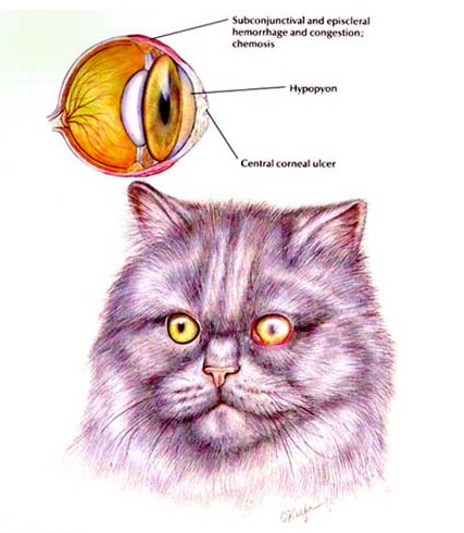
Signs of a Corneal Ulcer
- squinting or holding the eye closed due to pain
- excessive tearing
- light sensitivity
- rubbing the eye
- yellow-green mucus discharge from the eye
- clouding of the eye surface
- divot in the surface of the eye
Causes
The resulting defect can be superficial or deep, depending on the cause and whether infection develops within the tissue. Corneal ulcers are further classified as uncomplicated or complicated based on their healing.
Diagnosis
A corneal ulcer is diagnosed during an eye examination. The defect may be detected by visual inspection, but it requires the application of special stains to definitely determine the presence, size and the extent of the ulceration. Veterinary ophthalmologists also use equipment (slit lamp biomicroscope) to further magnify and inspect the tissues during an examination.
Depending on the severity, specimens may be collected for bacterial, fungal or viral culture and for cytological examination.
Once a corneal ulcer is diagnosed, it is further categorized as superficial or deep and complicated versus uncomplicated.
- Superficial corneal ulcer - as the name implies, just the outer layer of the corneal surface is damaged (much like a scrape on your skin).
- Deep corneal ulcer - in addition to the outer epithelial layer, the middle layer (stroma) is also damaged. Typically there is concern that infection is present in these types of ulcers, particularly if the ulcer started out as superficial and progresses to a deeper wound. The depth of the ulcer can progress to the worst case scenario - a descemetocele - in which the entire thickness of the cornea is damaged up to the final layer. An eye with a descemetocele is in great danger of rupturing and is considered a surgical emergency.
Corneal ulcers are also categorized based on the underlying, identifiable cause and their healing patterns.
- Non-complicated - these are ulcers that are usually due to trauma or another transient cause. Generally, they are superficial wounds that heal in an expected time frame of just a few days (from as few as 3 to 5 days upwards to 14 days).
- Complicated - these are corneal ulcers that are due to another identifiable underlying cause, such as an anatomic abnormality, and which may not heal unless the other issue is first succesfully addressed.
Another type of complicated corneal ulcer is the Refractory or Indolent Corneal Ulcer, in which an inherent abnormality within the corneal tissue itself is the root of the problem.
The healing of complicated ulcers can be prolonged.
Treatment
If an underlying cause for the ulceration or concurrent medical problem is identified, then those issues should be addressed to faciliate healing of the corneal ulceration. In addition, the following are necessary to prevent or treat infection and pain, and promote healing of the tissue:
- Infection
Corneal ulcers are treated with antibiotics, antifungals and / or antivirals depending on the diagnosis. Topical drops or ointments may be prescribed along with oral medications. Compliance with the recommended frequency of administration of these drugs is critical to their effectiveness in preventing or treating infection.
- Pain
The source of pain associated with corneal ulcers may be one of two places. With corneal ulceration, there may be concurrent spasm of internal eye muscles which also cause the pupil to constrict. By relaxing these muscles, with a medication that dilates the pupil, the pain associated with the spasms is alleviated.
With corneal ulceration there is also exposure of nerve endings in the tissue. These nerve endings are very sensitive and exposure of them results in pain. In some instances, depending on the type of the corneal ulceration, a therapeutic soft contact or bandage lens may be prescribed.
Applying a warm, moist compress, such as a wash cloth, over the affected eye for 5 to 10 minutes, two to three times a day, may provide some comfort.
- Promote healing
In a healthy eye, a superficial, uncomplicated corneal ulcer heals within a matter of days by the normal healing mechanisms of the tissue to injury. Preventing the animal from rubbing its eye or face against the floor or furniture is important to keep it from "undoing" what the body does to heal itself. An Elizabethan collar should be placed to prevent the animal from rubbing at its eye. Ophthalmologists may occasionally prescribe medications of various types to stimulate healing of the tissue.
In the case of a deep corneal ulcer or descemetocele, surgery may be the best option at promoting healing and preventing the eye from perforating. Depending on the ulceration or surgeon preference, the surgical procedures performed include conjunctival grafts, corneoscleral transpositions or corneal transplant.
Prognosis
The prognosis for superficial, uncomplicated corneal ulcers is good with appropriate treatment.
Treatment of complicated corneal ulcers, including successful treatment or management of the identified underlying cause, also carries a good prognosis.
The biggest risk is carried in pets with infected or deep corneal ulcerations. If not aggressively treated, these corneal ulcers can threaten the integrity of the eye and progress to perforation or rupture of the globe. Therefore, they pose a risk for loss of vision or the eye altogether. In cases of deep corneal ulcers or corneal perforation, surgery to prevent or repair the perforation may be required to save the eye.
Illustration reprinted with permission by the copyright owner, Hill’s Pet Nutrition, Inc.