
Forums
Guidance, support and wisdom to benefit and maximize the life and longevity of animals.
with a password reset link.
Be sure to check your SPAM folder if it's not in your inbox
Can't find it?
Need Help? Contact Us
Guidance, support and wisdom to benefit and maximize the life and longevity of animals.

VetVine Client Care

This is a photograph of the left eye of an 11 year old, MC, Labrador mix:
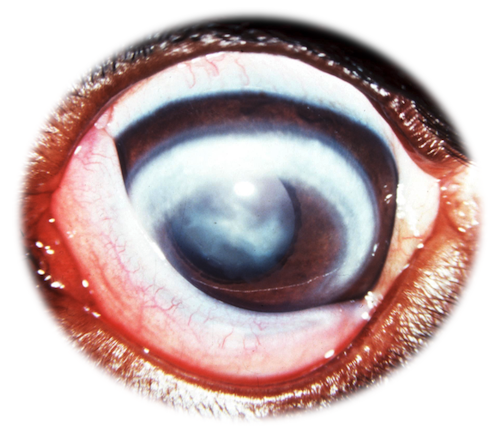
History: The owners noticed progressive cloudiness of the left eye over the past 6 months. The right eye was enucleated due to an intraocular iridociliary epithelioma and secondary glaucoma approximately 6 months prior. Vision seemed appropriate and the dog appeared comfortable.
Clinical Exam: Ophthalmic diagnostics were all normal (STT = 23 mm/min; IOP = 11 mm Hg; fluorescein stain negative).
All of the pathology that we see is located in the cornea. Of the 7 colors that we can see with corneal pathology (e.g. red (blood vessels), blue (corneal edema), yellow-green (pus), sparkly white (mineral), grey (fibrosis), creamy white (lipid), and brown (pigment)), this is creamy white.
1) What is your clinical diagnosis?
2) What other diagnostic tests should be completed?
Abnormal mineral and/or lipid in the cornea is consistent with corneal degeneration. This must be differentiated from corneal dystrophy. Corneal dystrophy is a suspected inherited condition in which there is mineral accumulation in the central cornea, usually in the subepithelial layer. As opposed to corneal degeneration, corneal dystrophy tends to be bilateral, ovoid to elliptical, non-progressive, non-inflammatory, does not affect vision, and occurs between 2-5 years of age.
In the this case, the lipid is present in the axial cornea as well as in an arc along the superior quadrant. This has been termed lipid keratopathy and, in people, arcus lipoides.
Lipid keratopathy is suggestive of metabolic disease and therefore a full medical work-up is indicated. Testing should include general screening (CBC, serum biochemistry) with special attention to cholesterol, trigylcerides, and ionized calcium as well as a thyroid level and possibly an ACTH stim if the serum chemistry suggests hyperadrenocorticism. The pet be fasted (12 hrs) prior to collecting samples to assess triglycerides, etc. when doing the work-up.
This dog was diagnosed with hypothyroidism and this was the cause of the lipid accumulation in his cornea.
The prognosis is fair. Treatment of the underlying hypothyroidism in this case prevented further lipid accumulation and the lesion did substantially fade over time, although residual stromal lipid remained present long-term.
If the lipid did not fade and a vision deficit was present, we would consider surgery (superficial keratectomy) to remove the abnormal tissue. However, the lipid could recur and therefore careful monitoring of the thyroid level and dog's lipid levels would be indicated.
This case was originally contributed by Brad Holmberg, DVM, MS PhD, DACVO (in May 2012)