Forums » Feline Medicine

"Shadow" - Not himself and eating less

  • February 7, 2014 2:06 PM EST

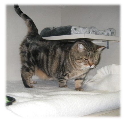
    "Shadow," is a long-time patient of mine and is examined regularly.  


    Signalment:  12 year old, MN, brown tabby DSH.

    Past History:  

    - Mild constipation - well controlled

    - Mild recurrent URTD


    Diet:  Science Diet Hairball Light


    "Shadow" presented to me recently with the complaint that he was "not himself" and he was eating less.  No report of vomiting, diarrhea or PU/PD.


    Physical Exam findings were:

    • BCS = 4/9
    • Weight = 5.77 kg (10 months prior he weighed 6.1 kg - this is 5.4% weight loss)
    • Grade I gingivitis
    • *Cranial abdominal mass noted
    • He was also a bit fractitious during the exam!

    Question:  What is Shadow's problem list and what are your rule outs at this point?
  • February 11, 2014 11:04 AM EST

    Summarizing Shadow's problem list:

    • 5% weight loss
    • Partial anorexia
    • Cranial abdominal mass
    • Pain
    • Gingivitis

    As for rule outs:
     
    Weight loss and partial anorexia are non-specific findings - can be caused by almost anything.
    Cranial abdominal mass - Could be involving the stomach, pancreas, liver, small intestine, lymph nodes, other?
    Don't forget about the pain - this is significant ...

    The initial diagnostic plan included collecting a minimum data base:
    • CBC = normal
    • Total T4 = normal
    • Urinalysis = normal
    • FeLV/FIV = negative
    • Chemistry Panel results:
     
    He was painful and fractious - so we gave him Buprenorphine 0.03 mg/kg transmucosally and then took survey abdominal radiographs.


    Question:  What do you see?
  • February 23, 2014 5:22 PM EST

    I think everyone can agree that the radiographs demonstrate an abdominal mass.


    Question:  Which organ is the abdominal mass involving?

    1)  Liver

    2)  Stomach

    3)  Pancreas


    Is there any other radiographic imaging you would consider to help you determine this?


  • February 24, 2014 3:26 PM EST

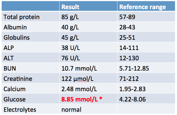
    There seems two choices from here in my view: either a contrast study of GIT or, as would more likely take place in my practice, an ultrasound.   It looks like liver to me but the liver enzymes (though there were only 2 reported) were within normal range. Any kind of change to liver parenchyma would usually provide some alteration to those values, i would think. 

  • February 27, 2014 11:37 AM EST

    Agreed regarding imaging options.  Although the following radiographic images are not from this case, here are some examples of how a positive contrast gastrogram can help to delineate abdominal masses / organ involvement (Reference:  Kristick KL et al, J Am Vet Med Assoc 236 (10), 2010).  


    So, the big question remains whether this cat has liver disease.  This brings up an important point - as demonstrated in this case:


    “Because marked hepatic disease can be present in patients with normal serum enzyme activity, finding normal values should not preclude further investigation.”  (Small animal internal medicine, ed 4)


    Reminder of alterations we can see in various tests in patients with hepatobiliary disease:


    With respect to liver function testing:


    •  Bilirubin, glucose, cholesterol, BUN, albumin


    – Hyperbilirubinemia is most sensitive/specific, esp. when >51 µmol/L (>3 mg/dL)


    •  Blood ammonia


    – Hepatic encephalopathy (signs would be clinically evident in affected patients)

    •  Serum bile acids (paired)
    –  Sensitive indicator in non-icteric cats
     
     Urine bile acids (4-8 hrs post-prandial)
    –  UBA/creat >4.4 = significant disease


    Question:  On the topic of hepatomegaly in the cat?  What are some rule outs to consider?

  • March 5, 2014 2:55 PM EST

    Here's my list of rule outs for hepatomegaly in cats.  Am I missing any others?


  • March 10, 2014 12:32 PM EDT

    We proceeded in our work-up.

    Because he was a little fractious and possibly painful it was appropriate to sedate him for the planned abdominal ultrasound (+/- biopsy).

    My choice for "Shadow" was:  Hydromorphone 0.1 mg/kg IM and Midazolam 0.2-0.4 mg/kg IM.


    On ultrasound, there was a liver mass (4.8 x 6.9 cm) with multiple anechoic areas noted.


    The list of rule outs for these findings included:

    • Hematoma
    • Abscess
    • Paracytic cyst
    • Biliary cyst
    • Tortuous biliary structures
    • Cystadenoma / adenocarcinoma
    • Polycystic liver disease
    • Metastatic liver disease

    Next for "Shadow?" ... we know that definitive diagnosis of most common feline liver diseases requires histopathologic examination of liver tissue.  Which of the following would you consider (and why)?
     
         FNA   -->   Tru-cut   -->   Laparoscopy   -->   Laparotomy    

  • March 14, 2014 3:59 PM EDT

    We know aspirates for cytology are far less likely to provide a definitive diagnosis. However, coagulation issues should be addressed if any of these options are considered. For the most comprehensive information, laparotomy or laparoscopy would be the most appropriate after coag and Vit K.

  • March 18, 2014 10:20 AM EDT

    Points to keep in mind regarding histopathologic examination of liver tissue:


    At least 6 portal areas are considered necessary to diagnose inflammatory disease

    (Requires ~15 mg tissue + 5 mg for culture).


    Vitamin K1 (2.5 mg/cat BID, SC, IM) is recommended for at least 24 hrs before biopsy.


    Regarding the options for obtaining samples:


    Fine needle aspirate

    • Inexpensive and easy to perform
    • Low risk, may only require sedation
    • Small sample size (3-5 mg) yield
    • Especially helpful for diagnosing lipidosis, lymphoma, and histoplasmosis
      ** However, per 
      Willard et al, J Feline Med Surg, 1999 - Misdiagnosis can occur as reported in this paper: 4 cats were diagnosed with lipidosis on FNA but, in fact, had infiltrative hepatic disease (3 had inflammatory disease and 1 had lymphoma)
    • Only ~50% agreement with histopathology

    Tru-cut biopsy
    • Requires general anesthesia
    • Risk of bleeding, fracture
    • Manual or semi-automatic devices only
    • Sample size yield is 15-20 mg (14g needle)

    Laparascopic biopsy
    • Limited invasiveness
    • Short recovery
    • Low risk
    • Large sample size yield - 45 mg
    • Can visualize and biopsy other organs
    • Can monitor for bleeding

    Surgical Biopsy
    • Allows for best visualization but is most invasive option
    • Allows for largest sample size and sampling multiple sites
    • Can monitor for bleeding