


The third eyelid or Nictitating Membrane is present in most animal species including dogs and cats. Associated with this third eyelid is a gland (gland of the third eyelid) which plays an important role in maintaining ocular health as it contributes to the production of tears. It produces 30 to 40 percent of the tear film of the eye. In its normal state the gland is not visible. However, when the gland becomes displaced from its normal position, it protrudes from behind the third eyelid and can appear as a pink to red bulbous mass. Because of this appearance, this prolapsed gland condition is commonly referred to as “cherry eye.” The problem is primarily seen in young dogs, including (among other breeds) the Cocker Spaniel, Lhasa Apso, Shih Tzu, Poodle, Beagle and Bulldog. The condition can also occur in cats including the Burmese. Although the exact cause is not known, there is a genetic predisposition to a weakness in the connective tissue elements that keep the gland normally positioned.
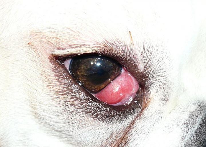 Photo Credit: Joel Mills; From Wikimedia Commons, the free media repository
Despite its appearance, cherry eye itself is not a painful condition nor is it usually considered an ocular emergency unless the animal rubs or scratches at the eye, demonstrates squinting (keeping the eye closed) and / or develops excessive discharge from the eye. In addition, the longer the tear gland is exposed, the more likely it will become irritated and inflamed. If the patient rubs at the eye, the gland can become swollen, bleed and develop a secondary infection. With prolonged exposure, the function of the tear gland can become compromised which can lead to serious complications including a dry eye condition. Therefore, surgery to repair the cherry eye (repositioning of the gland) is generally recommended.
There are several different surgical approaches to repositioning a prolapsed gland, and most of the techniques for cherry eye repair have a greater than 90 to 95 percent success rate. In some instances, however, a patient might require more than one attempt to achieve the desired outcome. Although surgical removal of the gland used to be commonplace years ago, this is no longer the case, as patients that have the gland removed are at risk for developing KCS (Keratoconjunctivitis sicca or Dry Eye condition) later in life, which can be a serious problem.
After surgical repair, an Elizabethan collar is usually recommended to prevent the patient from rubbing the eye during the healing process. Topical antibiotic and / or anti-inflammatory drops or ointments may be prescribed. Complications are not common but can include excessive swelling, secondary infection and corneal ulceration.